Chairside Ultrasound – the new standard for Dento-Facial injections
Chairside Ultrasound – the new standard for
Dento-Facial injections
by Dr Myles Holt
BDS, LLM (Heath&Medical), MSc (Aesthetic Medicine), FIADFE
AADFA Director & Head Trainer
Medico-Legal Advisor
Honorary Lecturer, MSc Aesthetic Dentistry, King's College London
Dental Surgeon
Flying Blind
In almost every aspect of modern dental practice, clinical procedures are made safer and more effective through the use of routine imaging.
Whether it be digital or conventional film; OPG, bitewing, periapical or CBCT; the use of some reliable method of imaging is considered best practice for all manner of dental treatments, from implants and RCT, through to extractions and simple restorations. Indeed, the failure of a dental practitioner to properly utilize available imaging to aid in everything from patient screening and diagnosis; to actively guiding treatment delivery; and conducting appropriate post-operative evaluation and complication management, would be considered a substantial departure from accepted standards, exposing them to regulatory action and litigation.
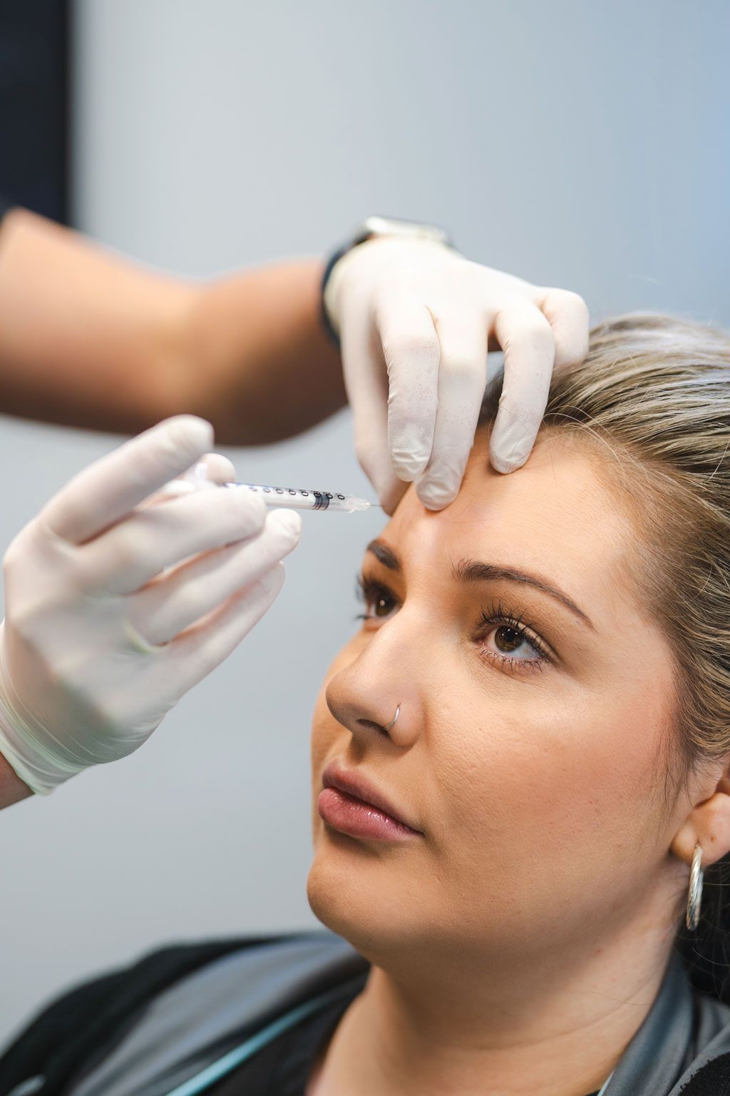
Essentially, it would be irresponsible and inappropriate to “fly blind” in traditional dentistry and indefensible if you did. Yet, until recently, a blind approach to treatment provision was the only option practitioners had when performing skin rejuvenation therapies; like Botox, Dermal Filler, Fat-dissolving injections and facial Thread Lifting; across the head and neck region and it had become the accepted norm.
So, while dental practitioners would never dream of blindly placing an implant into complex anatomy without the aid of imaging; running the risk of not only a poor result but serious complication if inadvertently encroaching on adjacent nervous or vascular structures; those placing Botox, dermal filler, and the like, simply had no other option. This is in large part due to the nature of the tissues being treated. Traditional dentistry, largely, works with hard tissue for which dental x-ray has been around for more than 100 years, with technology developing significantly over time to provide imaging tools of better quality, lower cost, and improved ease of use chairside. Facial soft tissues have been much harder to image directly by treating clinicians with technologies like MRI and CT having only been developed over a much shorter period which, despite significant advances, are still expensive, cumbersome, and inconvenient.
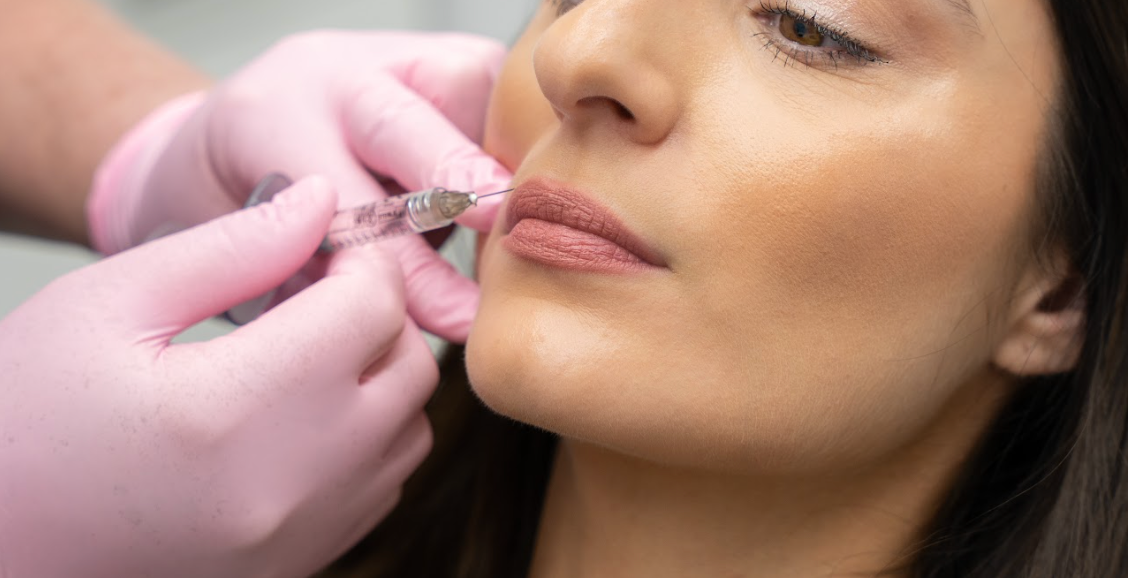
Yet, a critical aspect to many facial rejuvenation procedures (as with traditional dentistry), is correct anatomical placement. Whether it is depositing dermal filler into a strategic tissue space; aligning thread lifts within specific tissue planes; injecting Botox into individual muscle layers; placing fat-dissolving solution into designated fat compartments; or even achieving the correct depth of micro-needling penetration into the dermis; a failure to achieve correct anatomical orientation will reduce the effectiveness of treatment. More importantly, poorly planned, imprecise, and ill-conceived procedures, when working within the complex and closely related anatomy of the face, have the potential to cause serious complication, ranging from aesthetic asymmetries and irregularities; functional defects, if Botox impacts unintended muscles; through to skin necrosis and blindness if dermal filler inadvertently encroaches on vascular supply.
While practitioners tried to mitigate these risks by ensuring they had a comprehensive anatomical knowledge and deployed “safer” materials and techniques, differences in individual patient anatomy and practitioner ability meant that this was far from fool-proof. Ultimately it had to be accepted that flying blind was the norm when performing facial procedures and that even when practitioners were trying to do everything right, complications and poor outcomes could readily occur.
This has been particularly difficult for those just starting out and trying to learn these procedures, as it required a substantial amount of blind faith, a steep learning curve and an inherent nervousness without the benefit and reassurance brought about by the kinds of accurate imaging and pre-screening they were accustomed to in traditional dentistry. Thankfully, this has recently changed with developments in Ultrasound (US) technology setting the new standard for care.

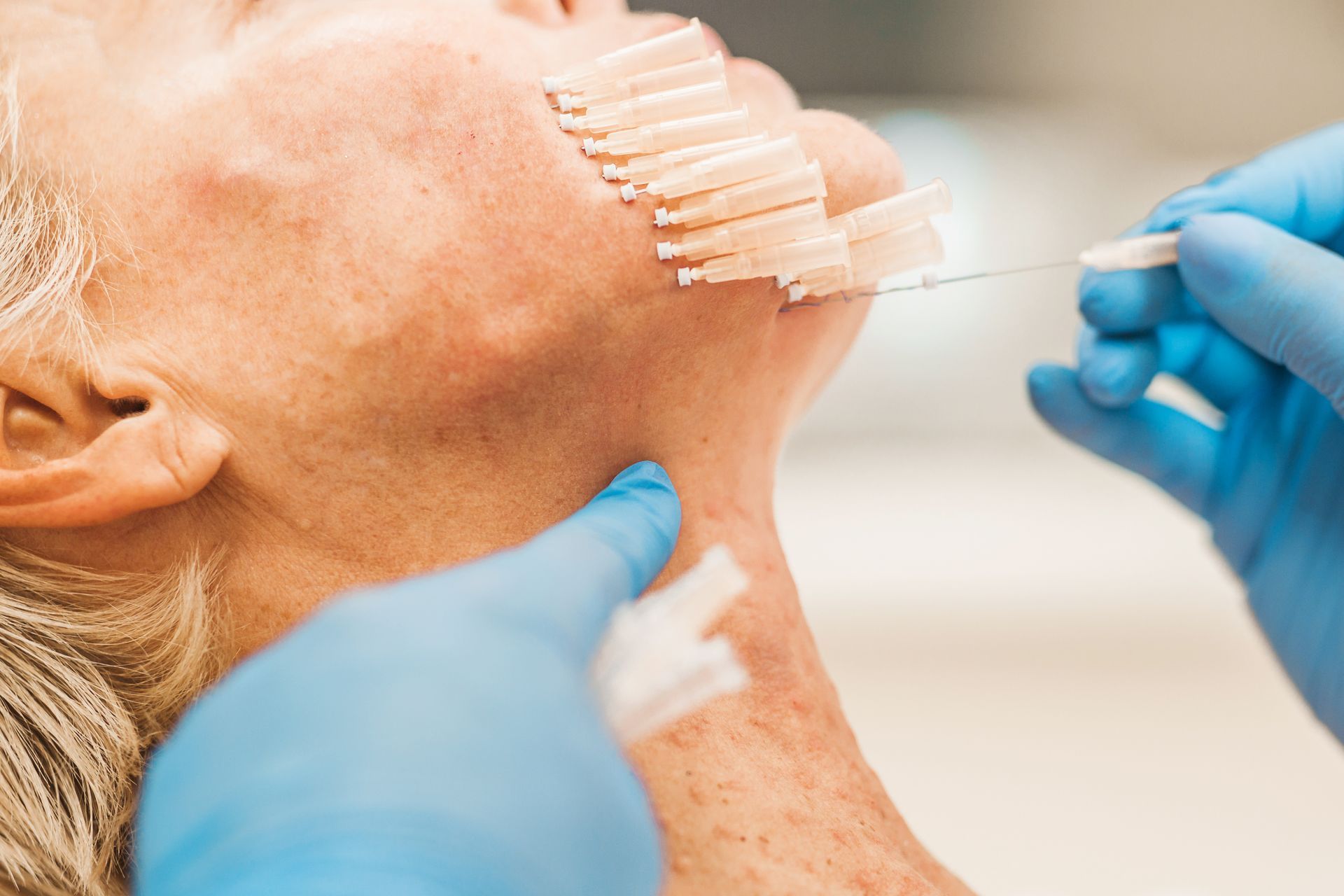
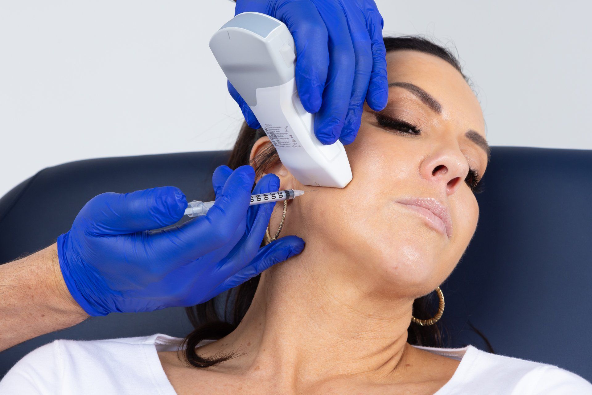
Above: Portable, wireless, hand-held, point-of-use Ultrasound technology, has set a new standard for patient care and practitioner education in dento-facial procedures.
Dentists lead the way
Through the pioneering work of the Australasian Academy of Dento-Facial Aesthetics (AADFA), the provision of facial rejuvenation treatments has become an accepted part of the practice of dentistry over the past decade. Additionally, Dentists are now recognised as among the very best practitioners to be offering these treatments to patients, with AADFA trained Dentists at the forefront of the industry, even contributing to the education of specialist plastic surgeons and dermatologists.
One of the main reasons for AADFA Dentists being able to readily move into this new area of practice and set the gold-standard for patient care in this field, is because of the clear parallels between the knowledge, skills and logistics needed to successfully deliver facial rejuvenation procedures with those required for traditional dental practice. The approaches seen in Dentistry, from high-level infection control and education, through to knowledge of material science and a focus on health-based responsible patient care, not cosmetic fads, have resulted in the bar being raised across the broader facial aesthetic community.
That trend now continues with AADFA becoming the first training organisation to incorporate the concept of active imaging, using the latest portable, wireless, hand-held, point-of-use US technology into patient assessment and treatment protocols for all facial rejuvenation therapies able to be performed by Dentists. The routine use of US has set the new standard for safety and efficacy when treating soft tissues, allowing Dentists to visualise precisely where target tissues lie; which areas to avoid; and to be guided in their treatment delivery.
This has not only set a new standard for patient care, providing comfort and reassurance to practitioners and patients alike, but it has set a new standard in the education of Dentists. Practitioners now learning dento-facial procedures can do so confidently, utilising the latest technology with AADFA’s clear parameters and clinical structure to flatten the learning curve and accelerate proficiency, all while increasing safety.
How is Ultrasound used?
Portable, point-of-use US technology utilises the same technology as all other ultrasound devices, with the advantage that it is low-cost, wireless, hand-held and can deliver real-time, high-quality diagnostic images direct to a tablet or smartphone, both quickly and painlessly, at chairside.
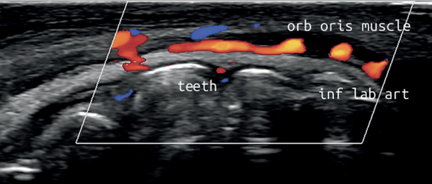
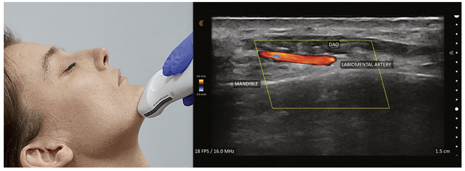
The US device emits a high-frequency sound wave which can penetrate several centimetres below the surface of the skin, where it is reflected differently by the various tissues being examined. This produces a live image of different densities on a grey-scale – bone appears different to muscle, which looks different to fat, which is distinguishable from the dermis and so on. Additionally, special settings allow for the colourful visualisation of not only the location of blood vessels, but also their size and velocity of flow.
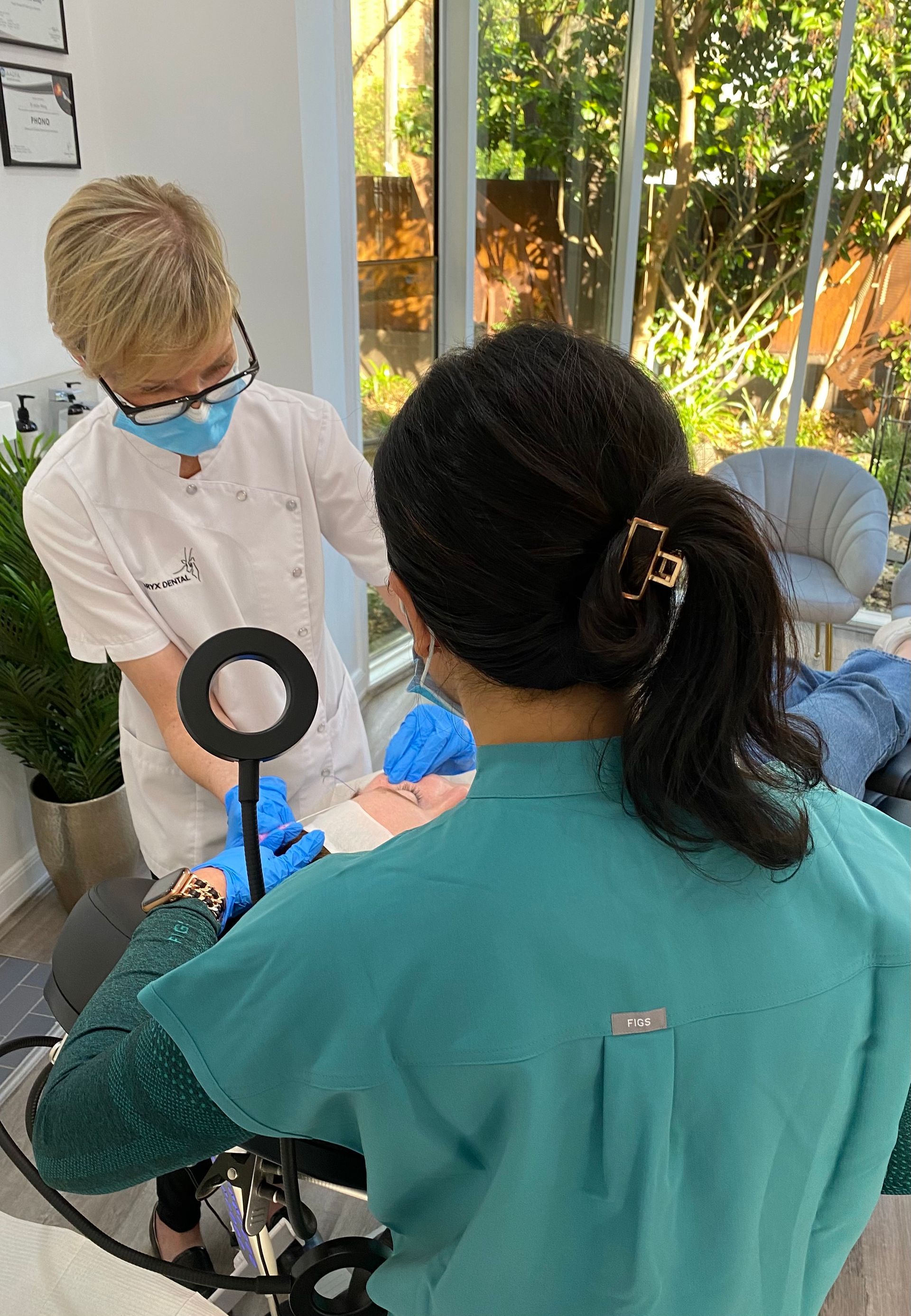
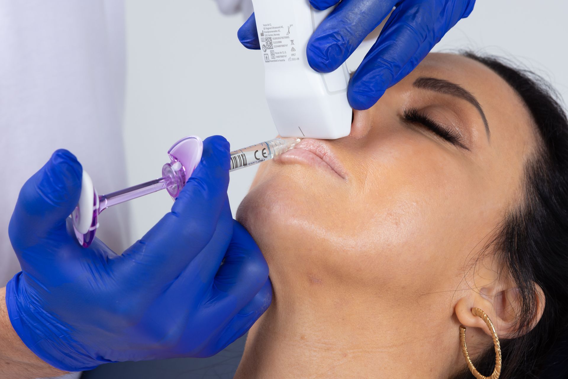
Above: Hand-held, chairside Ultrasound allows practitioners to visualise vital structures in real-time, improving treatment safety and efficacy.
Clinical research has demonstrated repeatedly that these unique imaging features allow practitioners to set new standards for safety and efficacy in three key ways:
Pre-screening & Diagnosis
US can be used in much the same way as traditional dental x-rays are utilised in consultations to visualise individual anatomy, plan appropriate treatment, and identify areas of risk. US allows us to develop a clear understanding of the face we are working with, mapping out key structures in advance, to plan treatment delivery in a manner which achieves the best results while actively preventing adverse events. The position, orientation, size, and depth of features can all be noted in advance, allowing a plan to be developed to target specific tissue layers with the likes of threads; avoid critical blood vessels with dermal filler; inject only certain muscles strategically with Botox; and even measure exact skin thickness so micro-needles penetrate precisely into the desired level of the dermis for optimum collagen stimulation.
Guided Treatment Delivery
US enables the clinician to perform guided injections for treatments involving Botox, Dermal Filler, fat-dissolving injections, and even facial thread lifts. In real-time practitioners can visualise their needle penetrating through the different layers of tissue until the desired treatment area and depth is reached. They can watch as the treatment is administered deep within the face, all of which previously would have to be done blindly, by feel, while hoping for the best.
Identification & Management of Complications
US enables practitioners to closely examine areas treated previously that may have experienced complications, diagnose them properly and deliver the appropriate treatment. Prime examples of this are complications arising from the injection of dermal filler.
US examination will quickly and easily reveal exactly where filler has been placed in the past and it is even possible to tell what type of product has been used from its unique echo signature. It would be possible to see if the filler is blocking a blood vessel and precisely where that blockage has occurred, which without active and accurate treatment, may lead to skin necrosis.
Thankfully, the best modern day dermal filler products consist of a Hyaluronic Acid gel and are therefore dissolvable with hyaluronidase enzymes. This allows the emergency treatment of dermal filler complications by dissolving the gel deposit and can be applied to resolve areas of lumpiness, irregularity or overfill, or importantly, to dissolve areas of filler that are causing a blockage of blood flow to the skin. However, the reversal agent is not without its problems, as it does not discriminate between natural hyaluronic acid in the skin (which forms a vital component adding hydration, elasticity, and integrity), and the filler product injected by the practitioner.
The problem is that previously, flying blind, practitioners had no way of knowing exactly where a blood vessel was blocked or precisely where an irregularity was positioned within the tissues. This meant that, in order to dissolve dermal filler to treat a complication, the only option was to “flood” a wide area of skin with the hyaluronidase, in the hope that it would reach the site required, wherever that may have been. Commonly, while emergency complications may have been addressed using this saturation approach, the skin is left in a compromised state - dry, flaky and irritated - requiring further intervention to restore health and aesthetics.
With the use of US, in the same way as injections of dermal filler can be guided in the first instance, so too can injections of hyaluronidase, such that only small droplets of the drug are needed, precisely placed in the exact area of vascular blockage or excess filler placement, avoiding collateral damage to adjacent skin and structures.
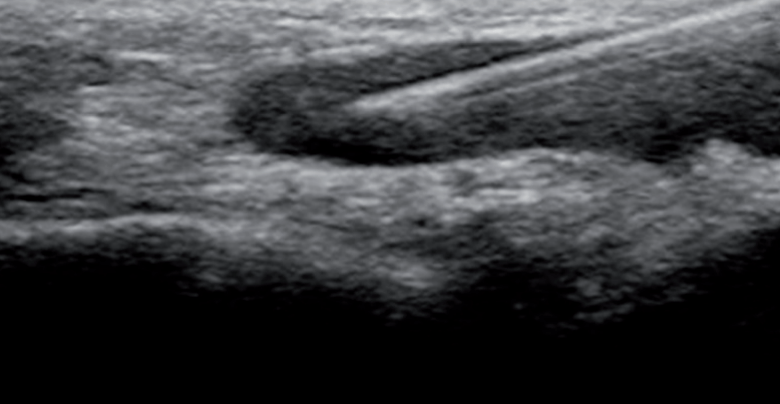
Above: Ultrasound can be used in real-time to guide injections and manage complications.
Adopt the new standard
A substantial body of research has shown that the ability to now use US chairside has set a new standard for clinical care in dento-facial rejuvenation, improving results and increasing safety.
With the availability of this imaging technology, facial procedures have now been brought into line with the long-standing norms of traditional dentistry. This may mean that should a practitioner fail to adopt this new standard of care, continuing instead to fly blind, they may very well be exposed to regulatory action and litigation in the event of sub-optimal outcomes or complications.
Adopting this new standard of care early will continue to set Dentists as the gold-standard in the industry. In a world where patients are increasingly moving away from practitioners who focus on an out-dated approach of basic “Cosmetic Injecting” in favour of responsible practitioners focused on proper healthcare delivery, (see article in the previous edition of Australasian Dentist magazine), they are now willing to pay a premium to ensure they receive a higher level of care. The use of US guided treatment gives practitioners a powerful and marketable point of difference.
Dentists starting their education in dento-facial procedures with AADFA, benefit from the fact that US technology protocols are now incorporated into every training program as standard. Those practitioners who have trained previously in dento-facial procedures will need to ensure they move swiftly to build US protocols in their practice, with AADFA now offering a bridging course called
“Phono”, designed to keep your practice at the forefront of modern dentistry.
For more information regarding all aspects of facial rejuvenation in Dentistry, contact the industry pioneers at AADFA HERE
PRESS PLAY ON THE VIDEO BELOW TO LEARN MORE